
🫀 When Adenosine Doesn’t Stop the Tachycardia – a 60-Year-Old Man with a Rapid Rhythm
- Faraz Afzal
- Oct 14
- 2 min read

🔎 Introduction
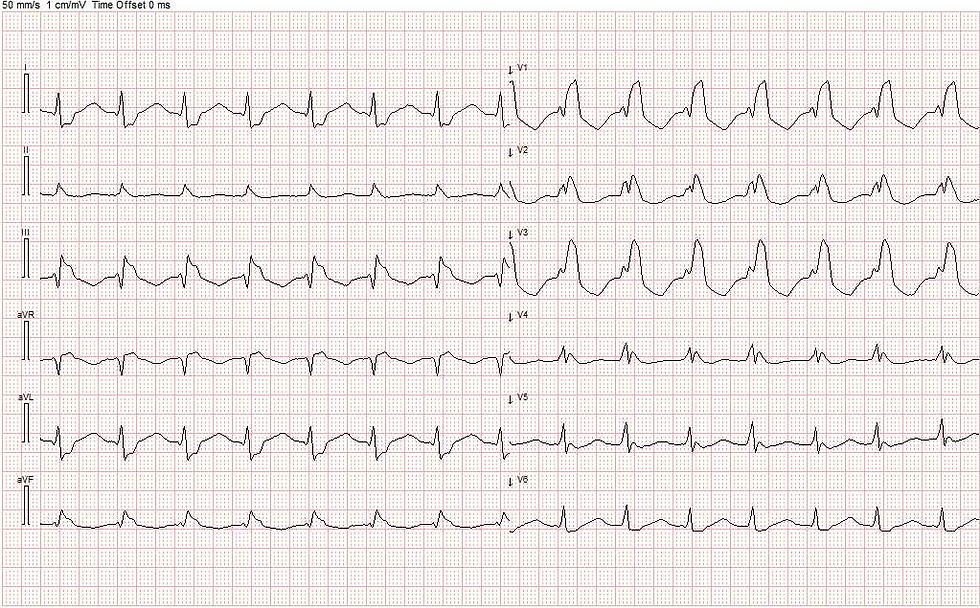
A 60-year-old man was admitted with palpitations, fever, and diarrhea. His ECG showed a rapid, regular rhythm at 182 bpm. Beta-blockers had no effect, and adenosine also failed to terminate the arrhythmia. So what was going on?
👨⚕️ Presentation and Initial Findings
The patient had a known right bundle branch block. On admission, the ECG revealed a regular, rapid rhythm at 182 bpm, with a broad QRS morphology unchanged from previous recordings.
Given these findings, a supraventricular tachycardia (SVT)—likely AVNRT or AVRT—was suspected. He was initially treated with a beta-blocker, but the heart rate remained unchanged.
💉 Adenosine Without Effect
A bolus of adenosine was administered to terminate the arrhythmia.Telemetry showed a brief AV block (pause in rhythm), followed by the immediate recurrence of the same tachycardia.
👉 This virtually rules out AVNRT or AVRT, since these types of reentry tachycardias are usually interrupted instantly by adenosine.


🧩 What’s Happening in the Heart?
Adenosine temporarily blocks conduction through the AV node.In atrial tachycardia (AT), however, the atria continuously generate impulses independently of the AV node.When adenosine is given, AV conduction stops briefly, but the atria keep firing, and as soon as the drug’s short half-life wears off, the tachycardia resumes.
➡️ The mechanism here is consistent with atrial tachycardia.
⚡ After Amiodarone – “Group Beatings”
The patient was then treated with amiodarone.Shortly after, he developed so-called “group beatings”—some beats dropped out, and the rhythm appeared in clusters.This pattern is classic for the Wenckebach phenomenon: a progressive prolongation of AV nodal conduction until a beat is blocked.
💡 The Wenckebach pattern further confirms the diagnosis of atrial tachycardia, as the atria continue to fire at a steady rate, while the AV node conducts only part of the impulses.

📉 Hemodynamic Consequences
Telemetry also recorded a photoplethysmogram (PPG) measured by pulse oximetry.The PPG reflects pulsatile changes in blood volume within the tissue during systole and diastole.When the heart contracts effectively and ejects blood into the arteries, local blood volume in the tissue (e.g., in the finger or earlobe) increases, producing a distinct peak on the pleth waveform.
In this case, the PPG clearly showed a reduction in pulse amplitude during tachycardia, illustrating how rapid atrial tachycardia negatively affects hemodynamics, with decreased stroke volume and reduced peripheral circulation.
⚡ Electrical Cardioversion
Since the arrhythmia persisted, the patient was electrically cardioverted to sinus rhythm.

🎯 Key Learning Points
✅ No effect of adenosine → argues against AVNRT/AVRT
✅ Wenckebach pattern after amiodarone → supports atrial tachycardia
✅ PPG changes → demonstrate hemodynamic impact - tachycardia is hymodynamically inefficient.
💬 Arrhythmia diagnosis often relies on evaluating several findings together. In this case, both the adenosine response and the Wenckebach pattern point toward atrial tachycardia


Comments